


Anatomical model of a cat's Scapula
Anatomical model of a cat's Scapula The model is intended for educational purposes, allowing you to practice various techniques for treating bone fractures. Thanks to the material and production technology, the model behaves like natural bone during drilling and cutting. Models can be disinfected with alcohol-based products, but cannot be steam sterilized as this will damage the model. Model size: 81 mm

Anatomical model of a cat's skull
Cat Skull A model prepared from a CT scan of a healthy cat. Ideal for educational purposes - both for veterinary students and for visual demonstration when communicating with clients.

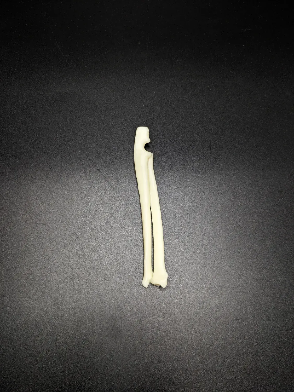
Anatomical model of the antebrachium
Anatomical model of the antebrachium of a cat The model is intended for educational purposes, allowing you to practice various techniques for treating bone fractures. Thanks to the material and production technology, the model behaves like natural bone during drilling and cutting. Models can be disinfected with alcohol-based products, but cannot be steam sterilized as this will damage the model. Model size: 124 mm


Anatomical model of the femur
Anatomical model of the femur of a dog The model is made using 3D printing technology. The model is intended for educational purposes and for practicing various bone fracture treatment techniques and can be used for preoperative plate contouring and for practicing fracture treatment techniques, as its material behaves similarly to natural bone during drilling and cutting. Models can be disinfected with alcohol-based products, but cannot be steam sterilized as this will damage the model. Model size - 156mm.

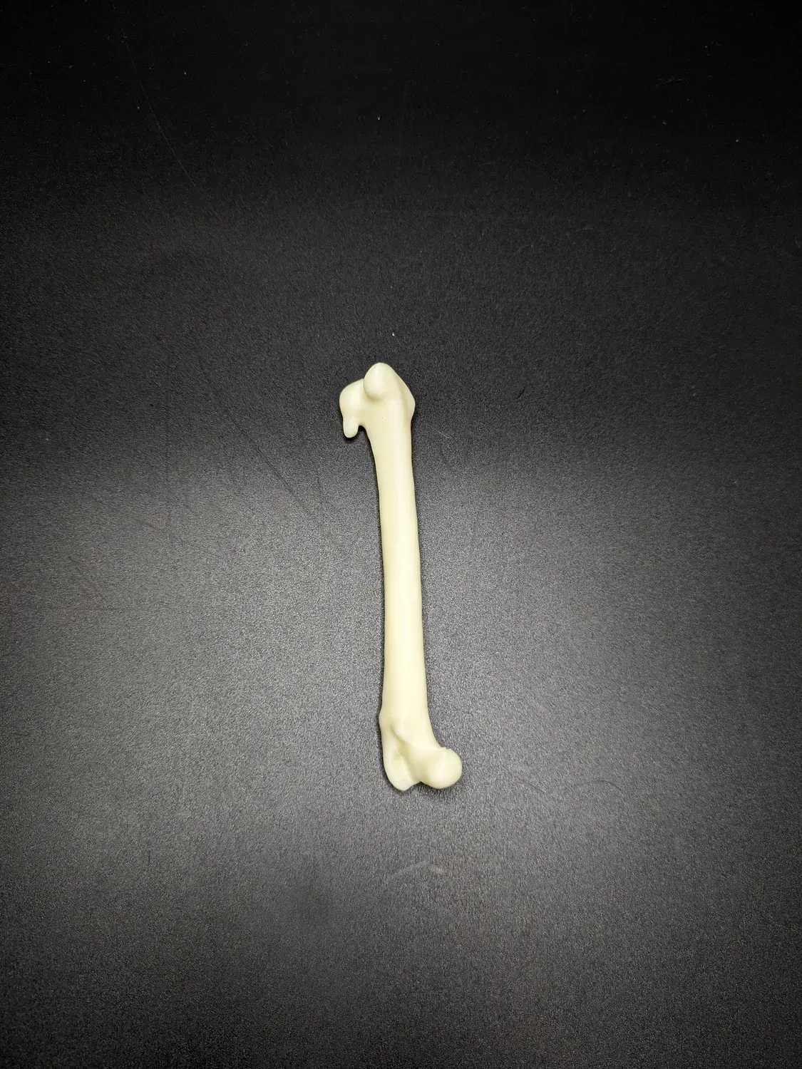
Anatomical model of the femur
Anatomical model of the cat's femur The model is intended for educational purposes, allowing you to practice various techniques for treating bone fractures. Thanks to the material and production technology, the model behaves like natural bone during drilling and cutting. Models can be disinfected with alcohol-based products, but cannot be steam sterilized as this will damage the model. Model size: 117 mm .


Anatomical model of the humerus bone
Anatomical model of the humerus of a dog The model is made using 3D printing technology. The model is intended for educational purposes and for practicing various bone fracture treatment techniques and can be used for preoperative plate contouring and for practicing fracture treatment techniques, as its material behaves similarly to natural bone during drilling and cutting. Models can be disinfected with alcohol-based products, but cannot be steam sterilized as this will damage the model. Model size - 145mm.
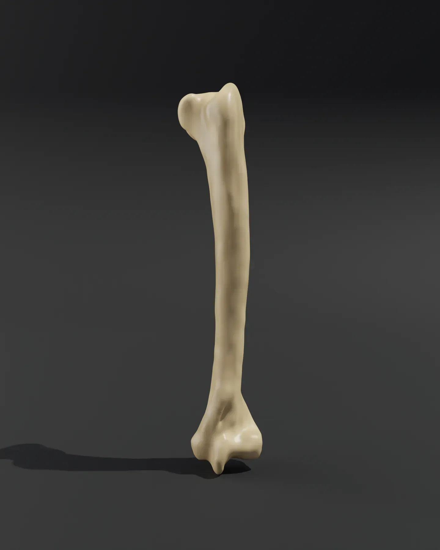
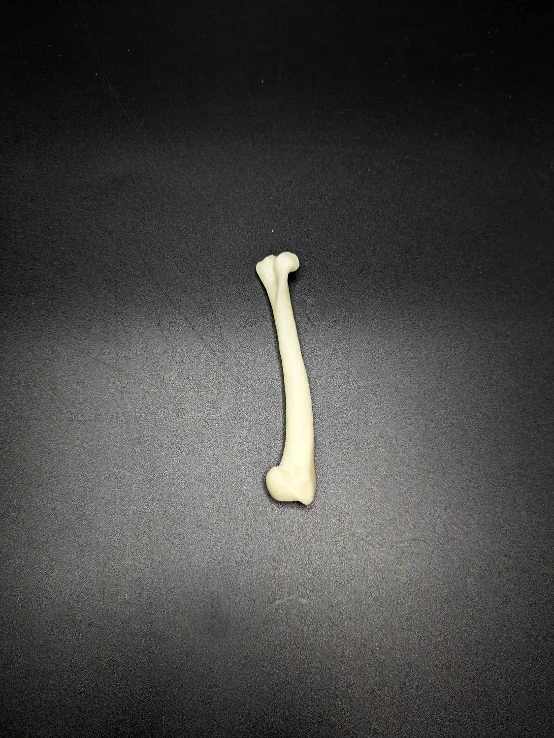
Anatomical model of the humerus bone
Anatomical model of the humerus of a cat The model is intended for educational purposes, allowing you to practice various techniques for treating bone fractures. Thanks to the material and production technology, the model behaves like natural bone during drilling and cutting. Models can be disinfected with alcohol-based products, but cannot be steam sterilized as this will damage the model. Model size: 106 mm
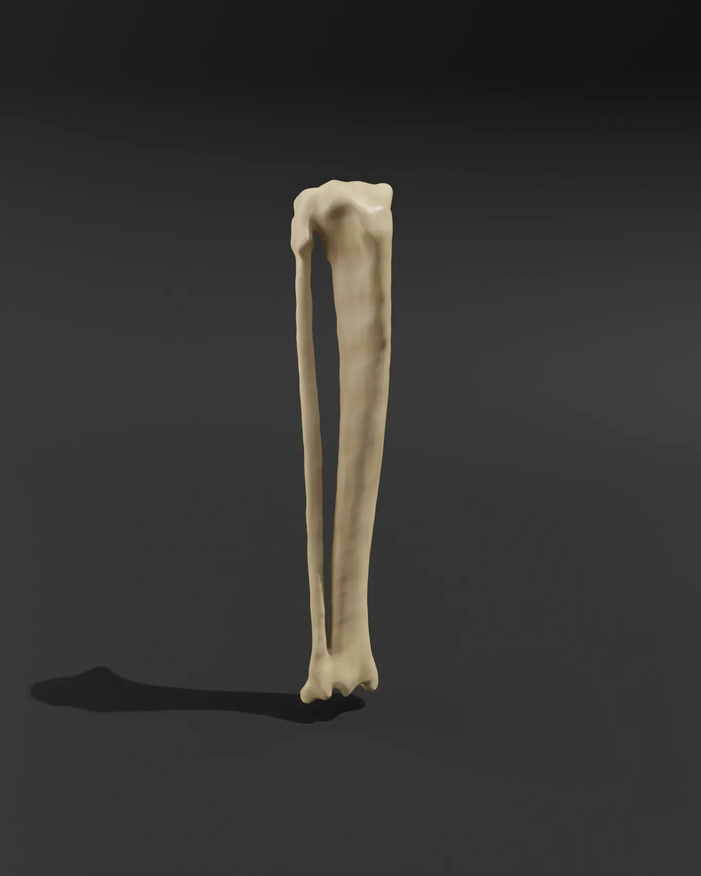
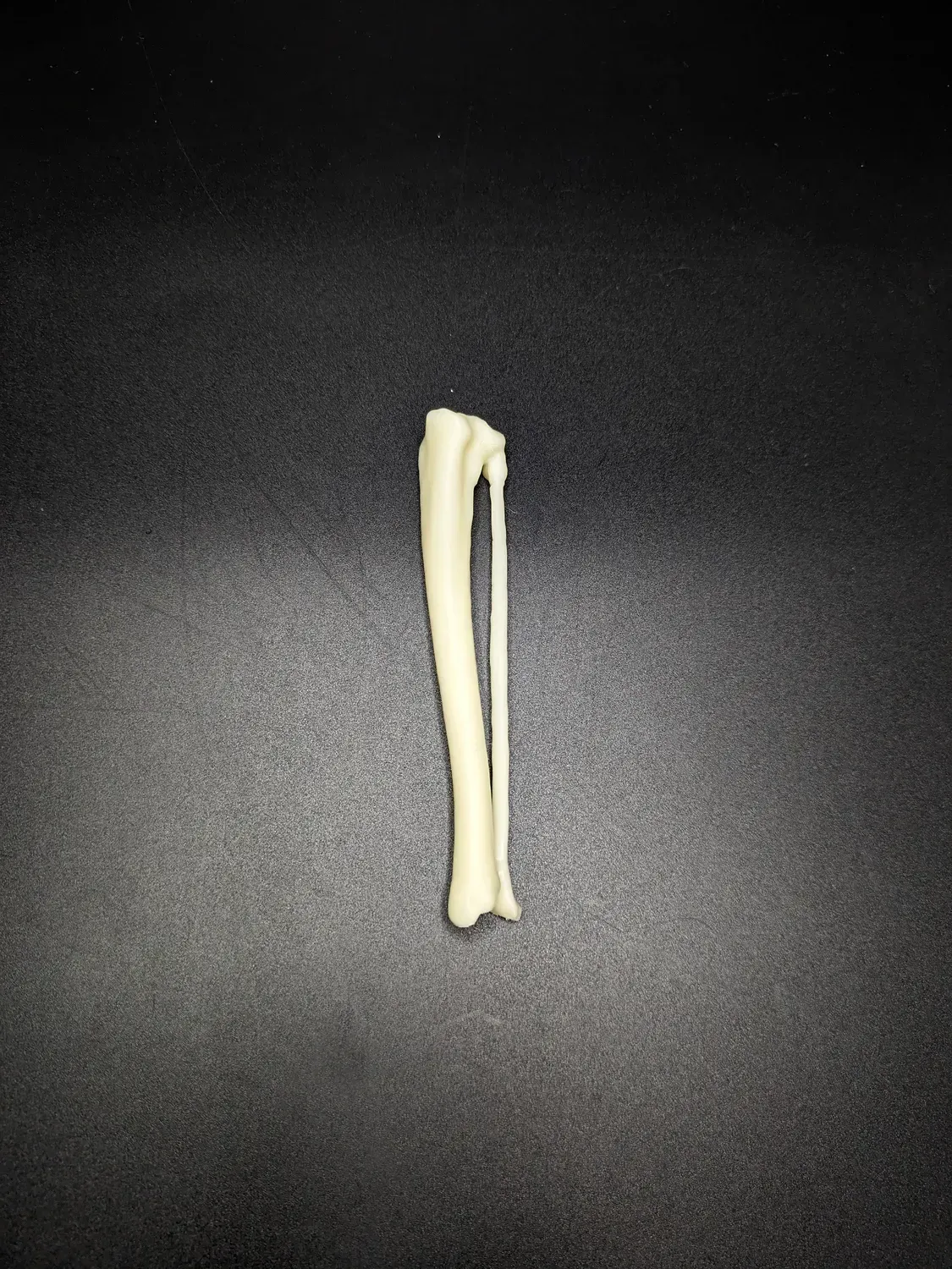
Anatomical model of the lower leg
Anatomical model of a cat's lower leg The model is intended for educational purposes, allowing you to practice various techniques for treating bone fractures. Thanks to the material and production technology, the model behaves like natural bone during drilling and cutting. Models can be disinfected with alcohol-based products, but cannot be steam sterilized as this will damage the model. Model size: 121 mm


Anatomical model of the pelvis
Anatomical model of the dog's pelvis The model is made using 3D printing technology. The model is intended for educational purposes and for practicing various bone fracture treatment techniques and can be used for preoperative plate contouring and for practicing fracture treatment techniques, as its material behaves similarly to natural bone during drilling and cutting. Models can be disinfected with alcohol-based products, but cannot be steam sterilized as this will damage the model. Model size - 133mm.


Anatomical model of the pelvis
Anatomical model of the cat's pelvis The model is intended for educational purposes, allowing you to practice various techniques for treating bone fractures. Thanks to the material and production technology, the model behaves like natural bone during drilling and cutting. Models can be disinfected with alcohol-based products, but cannot be steam sterilized as this will damage the model. Model size: 79 mm

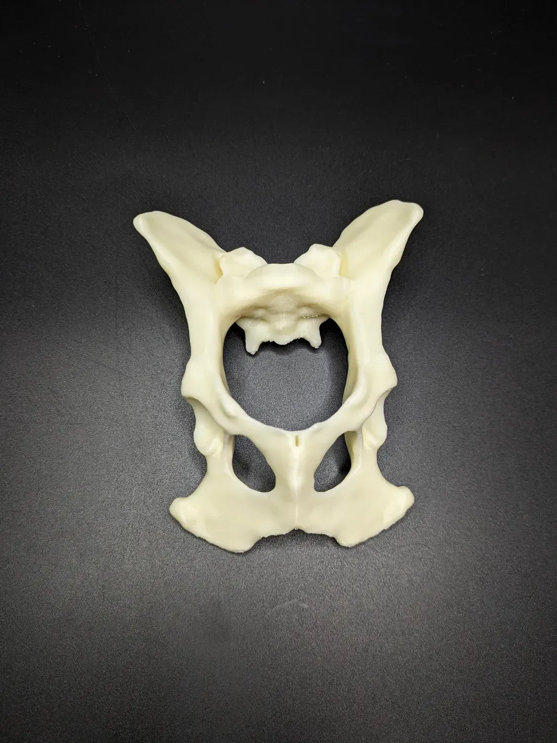
Anatomical model of the pelvis with coccyx
Anatomical model of the pelvis with a dog's tailbone The model is made using 3D printing technology. The model is intended for educational purposes and for practicing various bone fracture treatment techniques and can be used for preoperative plate contouring and for practicing fracture treatment techniques, as its material behaves similarly to natural bone during drilling and cutting. Models can be disinfected with alcohol-based products, but cannot be steam sterilized as this will damage the model. Model size - 133mm.

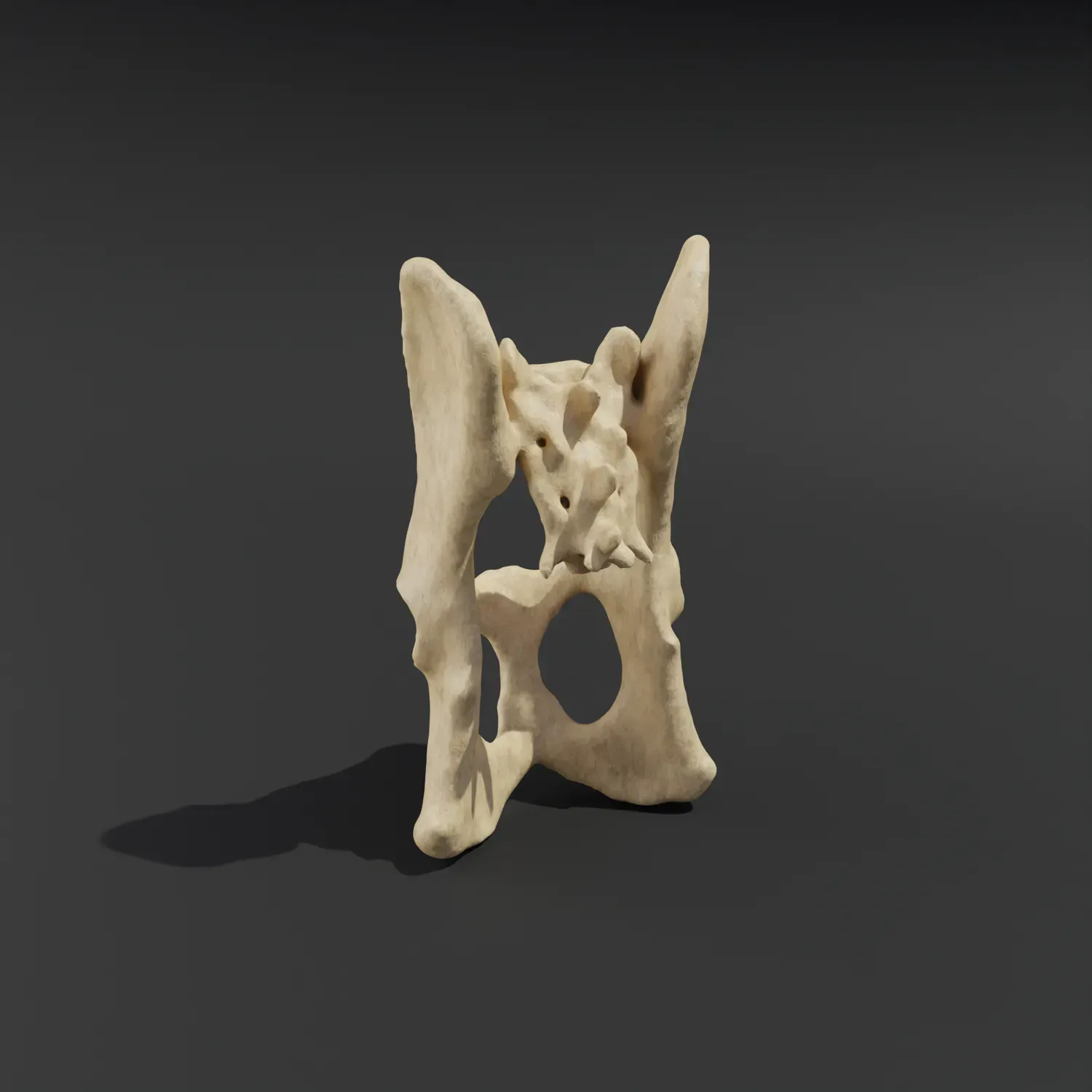
Anatomical model of the pelvis with coccyx
Anatomical model of the pelvis with a cat's tailbone The model is intended for educational purposes, allowing you to practice various techniques for treating bone fractures. Thanks to the material and production technology, the model behaves like natural bone during drilling and cutting. Models can be disinfected with alcohol-based products, but cannot be steam sterilized as this will damage the model. Model size: 79 mm .

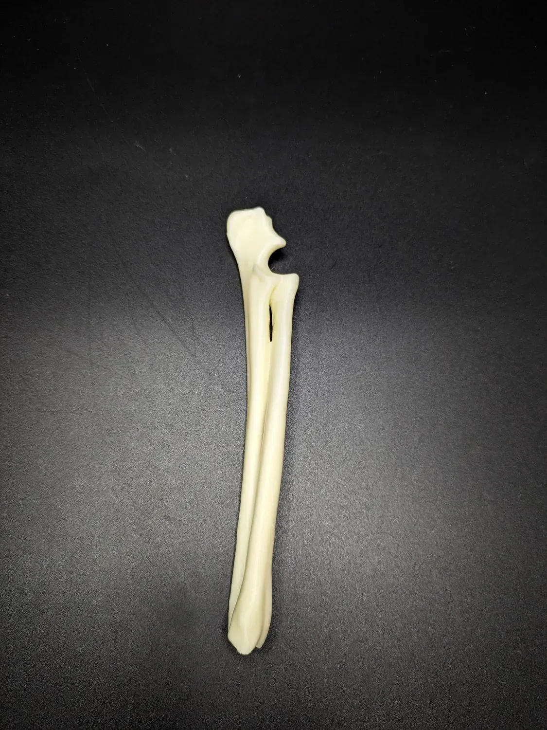
Anatomical model of the radius and ulna
Anatomical model of the radius and ulna of a dog The model is made using 3D printing technology. The model is intended for educational purposes and for practicing various bone fracture treatment techniques and can be used for preoperative plate contouring and for practicing fracture treatment techniques, as its material behaves similarly to natural bone during drilling and cutting. Models can be disinfected with alcohol-based products, but cannot be steam sterilized as this will damage the model. Model size - 173mm.


Anatomical model of the radius bone
Dog Radius Anatomical Model The model is made using 3D printing technology. The model is intended for educational purposes and for practicing various bone fracture treatment techniques and can be used for preoperative plate contouring and for practicing fracture treatment techniques, as its material behaves similarly to natural bone during drilling and cutting. Models can be disinfected with alcohol-based products, but cannot be steam sterilized as this will damage the model. Model size - 146mm.


Anatomical model of the scapula
Anatomical model of a dog's shoulder blade The model is made using 3D printing technology. The model is intended for educational purposes and for practicing various bone fracture treatment techniques and can be used for preoperative plate contouring and for practicing fracture treatment techniques, as its material behaves similarly to natural bone during drilling and cutting. Models can be disinfected with alcohol-based products, but cannot be steam sterilized as this will damage the model. Model size - 122mm.


Anatomical model of the tarsus joint
Anatomical model of the hock joint The model is made using 3D printing technology. The model is intended for educational purposes and for practicing various bone fracture treatment techniques and can be used for preoperative plate contouring and for practicing fracture treatment techniques, as its material behaves similarly to natural bone during drilling and cutting. Models can be disinfected with alcohol-based products, but cannot be steam sterilized as this will damage the model.


Anatomical model of the tibia and patellofemoral joint
Anatomical model of a dog's lower leg The model is made using 3D printing technology. The model is intended for educational purposes and for practicing various bone fracture treatment techniques and can be used for preoperative plate contouring and for practicing fracture treatment techniques, as its material behaves similarly to natural bone during drilling and cutting. Models can be disinfected with alcohol-based products, but cannot be steam sterilized as this will damage the model.


Anatomical model of the tibia bone
Anatomical model of the velo-humeral and fibular bones of a dog The model is made using 3D printing technology. The model is intended for educational purposes and for practicing various bone fracture treatment techniques and can be used for preoperative plate contouring and for practicing fracture treatment techniques, as its material behaves similarly to natural bone during drilling and cutting. Models can be disinfected with alcohol-based products, but cannot be steam sterilized as this will damage the model. Model size - 163mm.


Anatomical model of the ulna
Anatomical model of the dog's ulna The model is made using 3D printing technology. The model is intended for educational purposes and for practicing various bone fracture treatment techniques and can be used for preoperative plate contouring and for practicing fracture treatment techniques, as its material behaves similarly to natural bone during drilling and cutting. Models can be disinfected with alcohol-based products, but cannot be steam sterilized as this will damage the model. Model size - 173mm.


Anatomical model of the wrist joint
Anatomical model of the wrist joint of a dog The model is made using 3D printing technology. The model is intended for educational purposes and for practicing various bone fracture treatment techniques and can be used for preoperative plate contouring and for practicing fracture treatment techniques, as its material behaves similarly to natural bone during drilling and cutting. Models can be disinfected with alcohol-based products, but cannot be steam sterilized as this will damage the model.


Anatomical skull model - French Bulldog
French Bulldog Skull Model prepared from a CT scan of a healthy dog. Ideal for educational purposes - both for veterinary students and for visual demonstration when communicating with clients
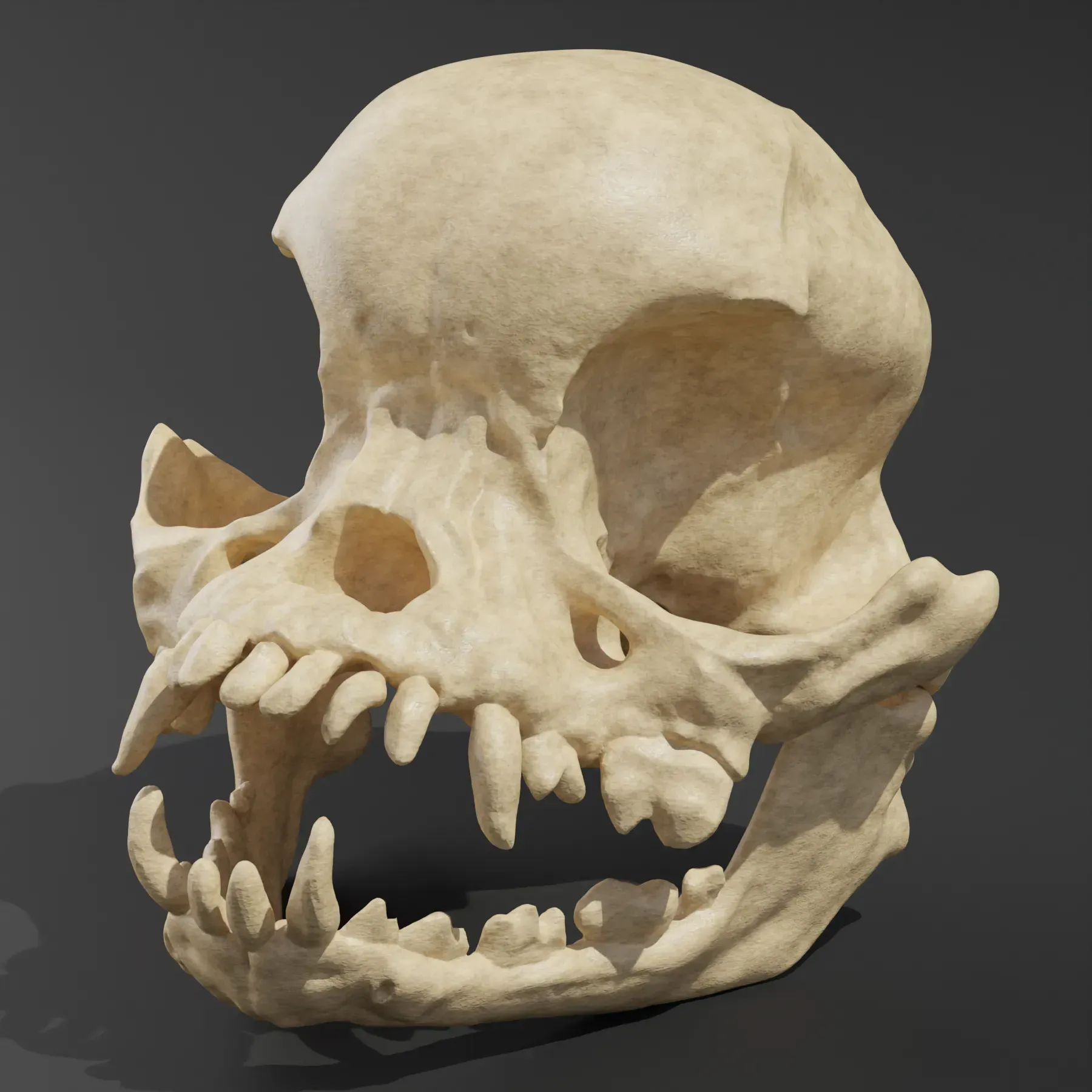
Anatomical skull model - Pug
Anatomical model of the Pug skull Model prepared from a CT scan of a healthy dog. Ideal for educational purposes - both for veterinary students and for visual demonstration when communicating with clients


Dismountable model. Anatomical model of the cervical spine of a cat
The model consists of 2 parts of interconnected vertebrae. Both parts are connected to each other thanks to magnets installed in the model. The model was prepared based on a CT scan of a healthy cat. Ideal for educational purposes - both for veterinary students and for doctors to explain in detail the issues of a clinical case to patient owners. The model can be subjected to cold sterilization and disinfection with special tools.


Dismountable model. Anatomical model of the cervical spine of a dog
The model consists of 2 parts of interconnected vertebrae. Both parts are connected to each other thanks to magnets installed in the model.
The model was prepared based on a CT scan of a healthy dog. Ideal for educational purposes - both for veterinary students and for doctors to explain in detail the issues of a clinical case to patient owners.
The model can be subjected to cold sterilization and disinfection with special tools.